This Website and Domain Name are for sale.

Squamous Cell Carcinoma

What is Squamous Cell Carcinoma?

Squamous cell carcinoma is a common skin cancer starting in squamous cells, making up the skin’s middle and outer layers. Usually not life-threatening, it can grow large if untreated, leading to serious complications. Most cases stem from ultraviolet (UV) radiation from sunlight or tanning devices.

Those who sunburn easily may find it in sun-exposed areas, while in people with Black and brown skin, it might appear in less exposed spots. Protection from UV light, such as sunscreen, can reduce the risk, highlighting the importance of vigilant skin care.

dermatologist-examining-skin-patient-with-dermatoscope

What are the symptoms of Squamous Cell Carcinoma?

Squamous cell carcinoma of the skin can manifest in various ways, often appearing on sun-exposed areas like the scalp, ears, or lips. However, it can occur anywhere, even inside the mouth or on the genitals.

The symptoms are noticeable and can include:

  • A firm bump, or nodule, that may match your skin color or appear pink, red, black, or brown.
  • A flat sore with a scaly crust that’s hard to ignore.
  • A new or raised area on an existing scar or sore, adding a twist to an old mark.
  • A rough patch on the lip that may turn into an open sore a warning sign you don’t want to miss.
  • A sore or rough area inside the mouth, a hidden but telltale sign.
  • A raised or wartlike sore on or in the anus or genitals, an unpleasant surprise.

These symptoms signal the body’s cry for attention, and it’s vital not to ignore them. If you notice any of these signs, consult a dermatologist.

What are the causes of Squamous Cell Carcinoma?

The root of squamous cell carcinoma lies in changes to the DNA within squamous cells, those skin cells with a crucial role. DNA is like a cellular instruction manual, dictating actions. Alterations prompt these cells to multiply swiftly, defying their natural lifecycle by surviving when they should perish.

This reckless growth leads to an excess of cells, which in turn can invade and wreak havoc on healthy tissue. Over time, these cells can detach, spreading their mischief to other corners of the body.

UV radiation takes the blame for most DNA alterations. Whether it’s sunlight, tanning beds, or those tanning lamps, UV radiation is the culprit. However, here’s an unexpected twist: skin cancers can sprout in areas not often exposed to sunlight. This suggests that other factors might join forces, increasing the risk. One such factor is having an immune system weakened by a specific condition.

How to diagnose Squamous Cell Carcinoma?

Squamous cell carcinoma has its roots in a mutation of the p53 gene. This gene is like the conductor of the cell division orchestra, instructing cells on when and how to replicate, replenishing themselves as needed. It’s a tumor suppressor, ensuring cell growth remains in check. However, this gene can go awry due to ultraviolet (UV) exposure from sunlight or indoor tanning beds.

When the p53 gene mutates, the cellular instructions get muddled. This miscommunication leads squamous cells to divide and reproduce excessively, birthing tumors—those unwelcome bumps, lumps, or lesions—in various nooks and crannies of the body. Think of it as a symphony conductor suddenly playing the wrong notes, leading to a discordant melody of cell growth and multiplication.

How to treat Squamous Cell Carcinoma?

When tackling squamous cell carcinoma, the goal is clear: remove the cancer from your body. Your treatment options hinge on factors like the cancer’s size, shape, and location.

Here’s a lineup of potential approaches:

  • Cryosurgery: Like freezing a pesky intruder, this method uses cold to destroy cancer cells.
  • Photodynamic therapy (PDT) is a light show for cancer cells. Blue light and light-sensitive agents team up to kick out unwanted guests.
  • Curettage and electrodesiccation: It’s like a double whammy. First, the cancerous lump gets scraped off with a spoon-like tool (curette). Then, an electric needle brings the heat, sealing the deal.
  • Excision: Time to cut it out. The cancer gets snipped from your skin, and then your skin gets stitched back together.
  • Mohs surgery: It’s all about precision. Layers of cancer-affected skin are peeled away, a technique often used for facial cancers.
  • Systemic chemotherapy: Heavy-duty medicines obliterate cancer cells across your body.

When it comes to medications, they’re not to be underestimated:

  • Skin creams with imiquimod or 5-fluorouracil work their magic on squamous cell carcinoma lounging in the top layer of your skin, giving them a proper eviction notice.
  • Cemiplimab-rwlc (Libtayo®): This immunotherapy superhero targets advanced forms of squamous cell carcinoma.
  • Pembrolizumab (Keytruda®): Another immunotherapy champion, tackling squamous cell carcinoma that surgery can’t handle.
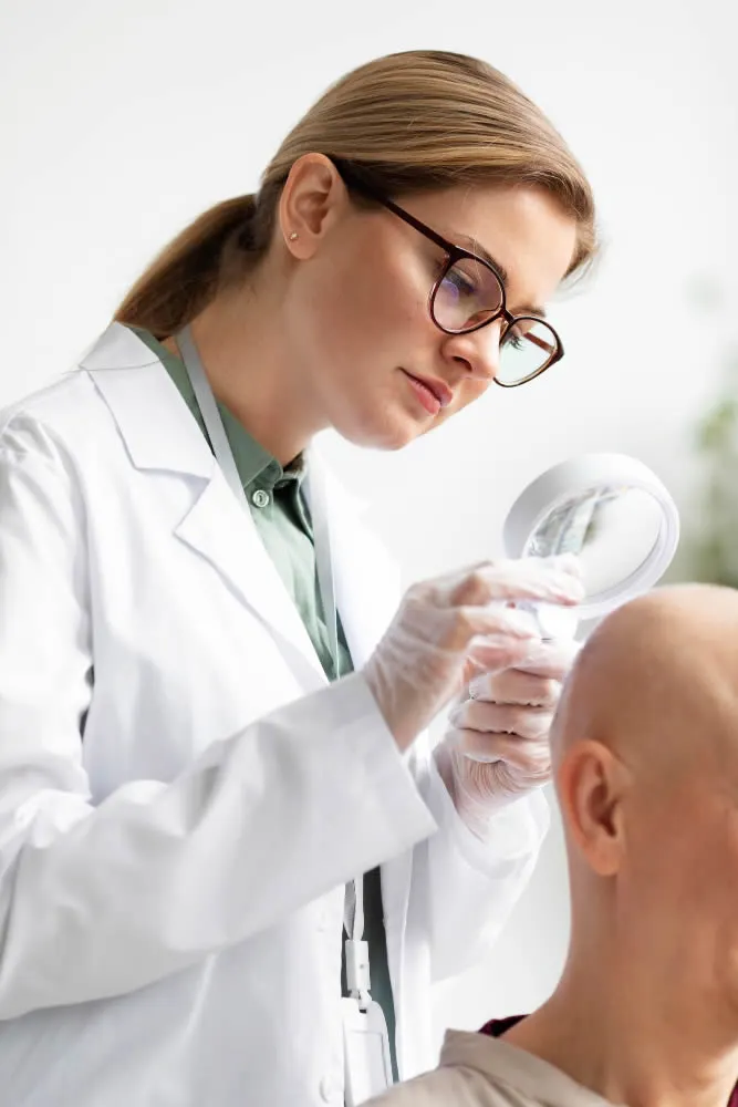
dermatologist-doing-check-patient-with-skin-cancer

FAQ About Squamous Cell Carcinoma

What are the side effects of the treatments for Squamous Cell Carcinoma?

The most common side effect of squamous cell carcinoma treatment is cosmetic changes to your skin, like scarring, after your healthcare provider removes the cancer from your body. If you take immunotherapy drugs to treat your cancer, talk to your healthcare provider about the side effects of the drugs.

How soon after treatment will I feel better?

The amount of time your body needs to heal after treatment varies for each person. The size, shape, and location also affect your healing time after treatment. On average, most people will recover within two to four weeks after treatment to remove cancer from their body. 

What can I expect if I have Squamous Cell Carcinoma?

Most cases of squamous cell carcinoma have a positive prognosis and an excellent survival rate if you receive an early diagnosis. Early detection and treatment prevent the tumor from growing and damaging other parts of your body. If your healthcare provider removes your cancer, there’s a chance it can return in the future. Make sure to follow up with your healthcare provider to verify you’re cancer-free. It’s also important to protect your skin from UV rays when outdoors.

What is Squamous Cell Carcinoma in situ?

Squamous cell carcinoma in situ is also known as Bowen disease. The term “in situ” means that the cancer cells are only in the top layer of your skin (epidermis). The most common places to find Bowen disease is on sun-exposed areas of your skin, but the condition can also appear on the skin near your anal cavity and genitals, like on your labia or vulva (vulvar cancer).

Is there a dermatologist near me in Chula Vista that offers treatment for Squamous Cell Carcinoma?

Yes. At our Chula Vista dermatology office we offer treatment for Squamous Cell Carcinoma to patients from Chula Vista and the surrounding area. Contact our office today to schedule an appointment.

Sebaceous Carcinoma

What is Sebaceous Carcinoma?

Sebaceous carcinoma, pronounced “suh-BAY-shus,” emerges as a rare skin cancer originating from oil-producing glands within the skin. These sebaceous glands, responsible for producing protective sebum, can be found extensively beneath the skin’s surface, especially in areas with hair.

While it predominantly targets the eyelids, sebaceous carcinoma can develop anywhere on the body. Its aggressive nature is evident through rapid spread, classifying it as metastatic cancer when it extends beyond the initial site.

Under various aliases like sebaceous gland carcinoma or meibomian gland carcinoma, it’s a formidable adversary seldom seen, affecting a mere 1% of eyelid tumors and presenting unique challenges in diagnosis and treatment.

dermatologist-examining-skin-patient-with-dermatoscope

What are the causes and risk factors of Sebaceous Carcinoma?

The precise triggers behind sebaceous carcinoma’s emergence unknown. Experts are still piecing together the puzzle of its origins. However, common threads are starting to emerge. Exposure to the sun’s ultraviolet rays, similar to other skin cancers, might play a role in its development. Notably, those who have undergone radiation therapy, particularly around the head or neck, could be more susceptible.

Certain factors that can make you more likely to get sebaceous carcinoma:

Muir-Torre Syndrome: People with a rare genetic condition called Muir-Torre syndrome have a higher risk of getting sebaceous carcinoma, as well as colorectal cancer.

Asian Descent: Some studies suggest that people of Asian descent might be more vulnerable to sebaceous carcinoma.

Age: Getting older can increase the risk.

Sun Exposure: Spending a lot of time in the sun without protection can also make it more likely.

Weakened Immune System: If your immune system is weakened due to conditions like cancer, HIV/AIDS, organ transplant medications, or drugs for arthritis or psoriasis, you are at a higher risk.

What are the symptoms of Sebaceous Carcinoma?

Advanced sebaceous carcinoma reveals a range of symptoms that warrant attention. Left untreated, this aggressive skin cancer can trigger various manifestations, including:

  • Loss of eyelashes, a concerning sign of its impact on the delicate eye area.
  • Oozing growths on both upper and lower eyelids, a distressing consequence of the disease’s progression.
  • Reddish eyes that resemble conjunctivitis, a symptom that can raise alarm.
  • Vision problems, a grave concern due to the potential impairment it poses.

How is Sebaceous Carcinoma treated?

The arsenal of treatments for sebaceous carcinoma encompasses diverse strategies tailored to the individual’s condition.

Surgical avenues take the forefront:

  • Wide Local Excision: A meticulous procedure that removes the tumor and a normal tissue margin. This helps ensure the cancer’s thorough elimination.

  • Mohs Surgery: Employing a staged approach, Mohs surgery incrementally removes the cancerous tumor and surrounding healthy tissue. During the procedure, thin slices of tissue are examined to gauge the need for further excision, guaranteeing complete eradication of cancer cells.

  • Radiation Therapy: Reserved for cases where surgery isn’t feasible, radiation therapy targets the cancer cells with high-energy rays, inhibiting their growth and division.

dermatologist-doing-check-patient-with-skin-cancer

FAQ about Sebaceous Carcinoma

What distinguishes Sebaceous Carcinoma from Sebaceous Hyperplasia and Sebaceous Cysts

Sebaceous hyperplasia is a benign growth within hair follicles, while sebaceous cysts are noncancerous growths forming beneath the skin due to clogged sebaceous glands.

When should I seek medical attention for skin growths or changes?

It’s advisable to consult a healthcare provider if you notice any unusual growths on your skin, especially around the eyes, or experience changes in moles or birthmarks.

What questions should I ask my dermatologist about Sebaceous Carcinoma?

Some relevant questions include understanding the causes, potential risks, suitable treatment options, and the need for cancer screenings based on your individual circumstances.

Is there a connection between hormonal changes and the development of Sebaceous Carcinoma?

While no direct link has been established, hormonal changes might influence the risk, but more research is needed for a conclusive answer.

Is there a dermatologist near me in Chula Vista that offers treatment for Sebaceous Carcinoma?

Yes. At our Chula Vista dermatology office we offer treatment for Sebaceous Carcinoma to patients from Chula Vista and the surrounding area. Contact our office today to schedule an appointment.

Microcystic Adnexal Carcinoma

What is Microcystic Adnexal Carcinoma?

Microcystic Adnexal Carcinoma is a rare cancer caused by an abnormal growth that starts in sweat glands. This condition typically shows up on the head and neck, especially in the middle part of the face. Sometimes it can appear in other places like the armpits, chest, private parts, and arms or legs, but this is pretty unusual. It looks like a scar but can be found on skin that’s been exposed to the sun.

The surprising thing is that this cancer doesn’t often spread to other parts of the body, which is good news. It’s rare by itself, and it can even get into bones, blood vessels, cartilage, muscles, and nerves. Even though it doesn’t usually spread, there’s still a risk that it might come back after treatment.

This cancer tends to follow along nerves in the body. It mostly affects older people. In rare cases it can affect kids.

dermatologist-examining-skin-patient-with-dermatoscope

Symptoms of Microcystic Adnexal Carcinoma

Skin Lesions: MAC typically presents as small, firm, skin-colored or pinkish lesions on the skin. They may resemble scars or small bumps. These lesions tend to grow slowly over time.

Slow-Growing Tumors: The tumors associated with MAC are generally slow-growing and painless, which means they may not cause discomfort in the early stages.

Infiltrative Nature: One characteristic of MAC is its infiltrative nature, which means it can spread along nerves, leading to numbness or tingling sensations in the affected area. This is known as perineural invasion.

Location: MAC often occurs on the head and neck, particularly in the central facial area. It can also appear in other areas of the body, although this is less common. The lesions are often found on sun-exposed skin.

Size and Shape: MAC lesions can vary in size but are generally small. They may be round or irregular in shape.

Absence of Symptoms: In some cases, MAC may not produce any noticeable symptoms, especially in its early stages. It may only be discovered during a routine skin examination.

Ulceration: In advanced stages, MAC lesions may become ulcerated or develop crusts.

Diagnosis of Microcystic Adnexal Carcinoma

  • Biopsy: A crucial starting point, a biopsy involves the careful extraction of a small tissue sample from the affected area. 
  • Microscopic Analysis: The extracted tissue undergoes rigorous microscopic testing. Skilled pathologists examine the cellular structure, looking for specific features indicative of MAC. This detailed analysis helps differentiate MAC from other conditions and confirms its presence.
  • Imaging Tests (Ultrasound and MRI): Ultrasound and MRI scans can also be used.

Treatment of Microcystic Adnexal Carcinoma

When addressing Microcystic Adnexal Carcinoma (MAC), a range of treatments is available, each tailored to the individual’s circumstances. These treatments encompass surgical interventions and therapeutic measures, with a focus on providing effective and personalized care:

  • Surgical Options: Small incisions and Mohs microsurgery serve as the cornerstones of surgical intervention. The distinctive advantage of Mohs micrographic surgery lies in its precision – it determines margins meticulously, reducing the need for repeated procedures and subsequent visits. In cases where Mohs micrographic surgery isn’t feasible, the surgical approach is guided by intraoperative frozen sections, ensuring accurate removal.
  • Follow-Up Care: Post-treatment, a critical aspect of MAC management, involves consistent follow-up visits. Patients are advised to consult their healthcare provider every six to twelve months. 
  • Addressing Lymph Node Metastasis: In severe cases of lymph node metastasis, surgical excision of affected lymph nodes may becomes  necessary. 
  • Chemotherapy Consideration: Chemotherapy may be used in conjunction with other therapies.
dermatologist-doing-check-patient-with-skin-cancer

FAQ about Microcystic Carcinoma

Is MAC contagious?

No, Microcystic Adnexal Carcinoma is not contagious. It is a type of skin cancer that develops internally and cannot be spread through contact.

Can MAC affect children?

While Microcystic Adnexal Carcinoma is more commonly observed in older individuals, it can occasionally affect children. However, this is quite rare.

What are the long-term effects of MAC treatment?

The long-term effects of MAC treatment can vary depending on the individual and the treatment methods used. Surgical procedures may leave scars, and chemotherapy might have side effects. Consulting with a healthcare professional can provide a clearer understanding of potential outcomes.

Is there a dermatologist near me in Chula Vista that offers treatment for Microcystic Adnexal Carcinoma ?

Yes. At our Chula Vista dermatology office, we offer treatment for Microcystic Adnexal Carcinoma to patients from Chula Vista and the surrounding area. Contact our office today to schedule an appointment.

Merkel Cell Carcinoma

What is Merkel Cell Carcinoma?

Merkel cell carcinoma is an uncommon skin cancer originating from Merkel cells in the skin’s top layer, often found in sun-exposed areas like the head, neck, arms, legs, and trunk. Also known as neuroendocrine carcinoma or trabecular cancer, it arises when Merkel cells grow uncontrollably.

Rapid growth and early spread to nearby lymph nodes, distant lymph nodes, skin, lungs, brain, bones, or organs are typical. These cells, positioned close to touch-sensing nerve endings, contribute to touch sensation.

Remarkably rare, Merkel cell carcinoma ranks as the second leading cause of skin cancer deaths, after melanoma.

dermatologist-examining-skin-patient-with-dermatoscope

What are the symptoms and causes of Merkel Cell Carcinoma?

Merkel Cell Carcinoma (MCC) often look like shiny or pearly lumps on sun-exposed areas like the face, neck, arms, and eyelids. These lumps can be skin-colored, red, purple, or bluish-red, and they may grow quickly. Darker-skinned individuals might find them on their legs, while younger people often notice them on the torso. These growths can even break open, forming wounds or sores that can be tender or itchy to the touch.

The primary contributor to MCC is exposure to ultraviolet (UV) rays from the sun or artificial sources like tanning beds. These UV rays can damage the DNA within skin cells, potentially leading to various skin cancers, including MCC. About 8 in 10 MCC patients also carry the Merkel cell polyomavirus (MCP), which usually remains dormant.

How to Diagnose Merkel Cell Carcinoma?

  • Physical Exam and Health History: The initial phase starts with a comprehensive physical examination with our dermatologist to gauge general health and identify any unusual signs, such as lumps or other irregularities. Reviewing the patient’s health history, including habits, past illnesses, and prior treatments, is crucial for establishing a complete picture.
  • Full-Body Skin Exam: The dermatologist will examine the skin’s entire surface, looking for unusual spots or bumps in color, size, shape, or texture. Additionally, the condition of lymph nodes, including their size, shape, and texture, is evaluated.
  • Skin Biopsy: If irregularities are detected, a skin biopsy is performed. This involves removing skin cells or tissues for microscopic analysis by a pathologist. The objective is to identify any signs of cancer within the sample.

Once MCC is confirmed, efforts shift toward understanding the extent of its spread, a process termed staging. Staging aids in determining the disease’s progression and tailoring suitable treatment approaches. Several tests and procedures contribute to this staging process:

  • CT Scan (CAT Scan): This imaging technique generates detailed images of the body’s internal areas from multiple angles. Dyes might be introduced to enhance organ visibility. CT scans can identify primary small cell lung cancer or locate MCC that has spread. They’re particularly useful for assessing the chest, abdomen, head, and neck regions.
  • PET Scan (Positron Emission Tomography Scan): A PET scan identifies malignant tumor cells in the body by injecting a small amount of radioactive glucose into a vein. The scan captures areas where glucose is utilized more actively, highlighting cancerous cells that exhibit heightened glucose consumption.
  • Lymph Node Biopsy: Various techniques are utilized to stage MCC through lymph node examination.
    • Sentinel Lymph Node Biopsy: The sentinel lymph node (the first node in a group) is removed during surgery. A radioactive substance or blue dye is injected near the tumor, guiding its flow to the lymph nodes.
    • Lymph Node Dissection: Surgical removal of lymph nodes allows for microscopic examination to identify signs of cancer. Regional or radical dissections are carried out based on the extent of lymph node involvement.
  • Core Needle Biopsy: A wide needle extracts a tissue sample for microscopic examination by a pathologist.
  • Fine-Needle Aspiration Biopsy: A thin needle retrieves a tissue sample for microscopic evaluation.
  • Immunohistochemistry: This laboratory test utilizes antibodies to detect specific antigens in tissue samples, aiding in diagnosing and differentiating various types of cancer.
dermatologist-doing-check-patient-with-skin-cancer

What are some of the treatments for Merkel Cell Carcinoma?

When it comes to treating Merkel Cell Carcinoma (MCC), there are several options depending on the patient’s needs. 

Treatment Options:

Surgery: Surgical intervention. Two main procedures are employed:

    • Wide local excision: This involves removing the cancerous growth along with surrounding tissue. Sometimes, a sentinel lymph node biopsy is performed simultaneously. If lymph nodes are affected, a lymph node dissection might follow.
    • Lymph node dissection: Removing lymph nodes to analyze them for cancer. Regional or radical dissections are conducted depending on the extent of lymph node involvement.

Radiation Therapy: High-energy radiation, like x-rays, is directed at the cancerous area to halt cell growth. External radiation therapy, which targets the body from outside, is a common approach. It may serve as palliative therapy to alleviate symptoms and enhance the patient’s quality of life.

Chemotherapy: Using drugs to halt cancer cell growth, chemotherapy can be administered orally, intravenously, or through injections. Systemic chemotherapy aims to reach cancer cells throughout the body, destroying them or stopping their division. 

FAQ About Merkel Cell Carcinoma

Are there stages of MCC, and how do they affect treatment?

Yes, MCC has stages that determine the extent of cancer spread. Staging guides treatment decisions, with localized MCC having better outcomes than metastatic cases.

What are the potential long-term effects of MCC treatment?

While treating MCC is crucial, it’s natural to wonder about potential long-term effects. Surgery, radiation, and chemotherapy may lead to side effects, including scarring, skin changes, and fatigue. These effects can vary depending on the treatment type and individual factors.

What factors influence MCC survival rates?

MCC survival rates are influenced by factors like overall health, age, and cancer stage. Localized MCC has a better prognosis compared to metastatic cases.

Is there a dermatologist near me in Chula Vista that offers treatment for Merkel Cell Carcinoma?

Yes. At our Chula Vista dermatology office, we offer treatment for Merkel Cell Carcinoma to patients from Chula Vista and the surrounding area. Contact our office today to schedule an appointment.

Melanoma

What is Melanoma?

Melanoma is a type of skin cancer originating from melanocytes, cells in the skin’s deep epidermal layer. These cells produce melanin, a pigment responsible for skin coloration, which also protects the skin from the harmful ultraviolet (UV) radiation of the sun.

Prolonged exposure to UV radiation can cause sunburn and damage the DNA in skin cells, potentially leading to uncontrolled cell growth and cancer. While melanoma is often associated with sun-exposed areas, it can also develop in moles or even in areas not typically exposed to the sun.

Detecting and treating melanoma early is crucial, as it can be aggressive and life-threatening if left unchecked.

dermatologist-examining-skin-patient-with-dermatoscope

Causes and genetic risk factors of Melanoma

Melanoma, a serious type of skin cancer, primarily from damage to the DNA of skin cells, due to exposure to ultraviolet (UV) radiation from the sun or tanning beds.

DNA damage can lead to uncontrolled cell growth, forming malignant tumors. While UV radiation is a significant environmental risk, genetic can also play a role in melanoma’s development.

Key causes and genetic factors include:

  • Ultraviolet (UV) Radiation: Prolonged and intense exposure to UV rays from natural sunlight or artificial sources significantly heightens melanoma risk.
  • Moles: Individuals with numerous or atypical moles (dysplastic nevi) are at a higher risk.
  • Skin Type: Fair-skinned individuals, especially those with light hair, blue or green eyes, and freckles, are more susceptible due to lower melanin levels, which offers some UV protection.
  • Family History: A person’s risk increases if a first-degree relative (parent, sibling, or child) has been diagnosed with melanoma.
  • Genetic Mutations: Specific gene mutations, such as those in the BRAF, NRAS, or CDKN2A genes, can predispose individuals to melanoma.
  • Previous Cancer Diagnosis: A history of melanoma or other skin cancers elevates the risk for future melanomas.
  • Weakened Immune System: Those with compromised immunity due to conditions like HIV/AIDS or treatments like organ transplantation are more vulnerable.

Prevention and regular skin checks are vital, given these risk factors, to detect and treat melanoma at its earliest stages.

Significance of early detection and symptoms

Early detection is important for melanoma, as timely intervention can greatly impact treatment outcomes and overall prognosis.

Detecting melanoma in its initial stages, before it spreads deeper into the skin or to other parts of the body, offers several crucial advantages:

  • Improved Treatment Success: Melanoma caught early is often highly treatable with various approaches, including surgical removal.
  • Minimized Risk of Metastasis: Early-stage melanomas are less likely to have spread to lymph nodes or distant organs, reducing the risk of metastatic disease.
  • Less Invasive Treatments: Smaller, localized melanomas are typically easier to remove surgically, resulting in less extensive procedures and quicker recovery times.
  • Preservation of Appearance: Early detection can potentially prevent the need for more extensive surgeries that might impact the appearance of the affected area.

Symptoms and self-Examinations:

Vigilance in self-examination and recognizing potential symptoms are key to early detection. Keep an eye out for the ABCDE characteristics of melanoma:

  • Asymmetry: One half of the mole or growth doesn’t match the other half.
  • Border: The edges are irregular, ragged, or blurred.
  • Color: The color varies within the mole or lesion.
  • Diameter: The size is larger than a pencil eraser (about 1/4 inch or 6mm).
  • Evolving: Any changes in size, shape, color, or other traits over time.

If any of these characteristics are observed, or if a mole or growth becomes itchy, painful, or starts bleeding, seeking prompt medical attention is crucial. Regular self-examinations, combined with professional dermatological check-ups, play a pivotal role in catching potential melanomas at their earliest, most treatable stages.

How to diagnose Melanoma at different stages?

Diagnosing melanoma involves distinct methods tailored to each stage of the disease.

  • Stage 0 (Melanoma In Situ): Diagnosis involves a skin biopsy. A small piece of the suspicious area is removed and examined under a microscope to confirm the presence of abnormal cells.
  • Stage I: Tumor size and thickness are assessed through biopsy. If there’s a concern about the cancer’s spread to nearby lymph nodes, a sentinel lymph node biopsy might be performed to determine its involvement.
  • Stage II: Biopsy evaluates tumor thickness, ulceration, and mitotic rate. Sometimes, a sentinel lymph node biopsy assesses potential lymph node involvement.
  • Stage III: Diagnosis encompasses evaluating regional lymph nodes and nearby skin for cancer spread. Imaging techniques like PET, CT, or MRI help identify distant metastasis.
  • Stage IV (Metastatic Melanoma): Diagnosis involves comprehensive imaging scans, including CT, PET, and MRI, to pinpoint the extent of the cancer’s spread. Biopsies of affected organs might be conducted to confirm the presence of melanoma.


Treatment approaches for Melanoma

The treatment path for melanoma depend on the cancer’s stage and the patient’s overall health. Surgery typically is the first option.

Factors like the size and location of the cancer determine the extent of healthy tissue removal. While surgical excision can often be performed under local anesthesia in a dermatologist’s office, more complex cases might require alternative treatments.

Melanoma treatment modalities:

  • Melanoma Surgery: In the early stages, surgical intervention holds strong potential for curing melanoma. Dermatologists perform this procedure in-office, numbing the skin with a local anesthetic before excising the melanoma and the surrounding margins of healthy skin.
  • Lymphadenectomy: Should melanoma spread, lymph nodes near the initial site of diagnosis might need removal. This preemptive measure can impede the cancer’s dissemination to other body areas.
  • Metastasectomy: Targeting small melanoma traces within organs, metastasectomy involves surgical removal to prevent further progression.
  • Targeted Cancer Therapy: Focused on specific cancer cells, targeted therapy employs drugs to attack them, sparing healthy cells. This precision approach minimizes collateral damage.
  • Radiation Therapy: Employing high-energy rays, radiation therapy targets cancer cells and shrinks tumors, providing a non-surgical avenue for treatment.
  • Immunotherapy: This treatment triggers the immune system to effectively combat cancer cells.
dermatologist-doing-check-patient-with-skin-cancer

FAQs About Melanoma

How common is Melanoma compared to other skin cancers?

While melanoma isn’t the most prevalent form of skin cancer, it is the most lethal. It accounts for a smaller percentage of skin cancer cases but a significant majority of skin cancer deaths.

Is Melanoma more common in certain geographical areas?

Melanoma tends to be more prevalent in areas closer to the equator and at higher altitudes due to increased UV radiation exposure.

Do sunscreen lotions guarantee protection against Melanoma?

While sunscreens significantly reduce UV exposure, they don’t provide complete protection. It’s essential to use them alongside other protective measures like wearing protective clothing and avoiding peak sun hours.

Is there a dermatologist near me in Chula Vista that offers treatment for Melanoma?

Yes. At our Chula Vista dermatology office we offers treatment for Melanoma to patients from Chula Vista and the surrounding area. Contact our office today to schedule an appointment.

Kaposi Sarcoma

What is Kaposi Sarcoma?

Kaposi sarcoma is a type of cancer that affects the skin and mucous membranes, which line various parts of the body including the gastrointestinal tract from the mouth to the anus. This cancer is characterized by the development of purple patches or nodules on the skin and mucous membranes. These growths can spread to lymph nodes and the lungs. Kaposi sarcoma is more commonly found in men and individuals with weakened immune systems.

dermatologist-examining-skin-patient-with-dermatoscope

What are the causes and risk factors for Kaposi Sarcoma?

Kaposi sarcoma is caused by an infection with human herpesvirus 8 (HHV-8), also known as Kaposi sarcoma-associated herpesvirus (KSHV). This virus is responsible for triggering the development of Kaposi sarcoma in individuals who are infected with it. HHV-8 is a member of the herpesvirus family, including viruses like herpes simplex virus and varicella-zoster virus.

Once a person becomes infected with HHV-8, the virus can lie dormant within the body for a long period without causing any symptoms. However, in some individuals, especially those with weakened immune systems, HHV-8 can reactivate and lead to the development of Kaposi sarcoma.

It’s important to note that not everyone who is infected with HHV-8 will develop Kaposi sarcoma. The interaction between the virus and the individual’s immune system and other factors plays a role in whether the cancer develops.

Certain populations are at higher risk for both HHV-8 infection and subsequently Kaposi sarcoma. These include people with suppressed immune systems (such as those with HIV/AIDS or those who have received organ transplants) and individuals living in regions where HHV-8 is more common.

What are the symptoms of Kaposi Sarcoma?

The symptoms of Kaposi sarcoma can vary depending on the location and extent of the cancer.

Common symptoms include:

  • Skin Lesions: Kaposi sarcoma often begins with the development of purplish or reddish patches on the skin. These patches can be flat or raised, appearing anywhere on the body. They can grow over time and become nodules or tumors.
  • Mucous Membrane Lesions: Lesions can also occur on the mucous membranes that line the mouth, nose, throat, anus, and genital areas. These lesions might cause discomfort, pain, or bleeding depending on their location.
  • Internal Lesions: Kaposi sarcoma can develop internally, affecting organs such as the lungs, gastrointestinal tract, and lymph nodes. Internal lesions can lead to symptoms such as difficulty breathing, coughing blood, abdominal pain, or bleeding.
  • Swelling: If the lymph nodes are affected, particularly those in the groin, it can result in painful swelling in the legs.

It’s important to remember that Kaposi sarcoma symptoms can vary widely from person to person. Some individuals may have only a few small skin lesions, while others might experience more widespread and severe symptoms.

Symptoms often depend on the specific type of Kaposi sarcoma, its stage, and the individual’s overall health. If you suspect you have symptoms of Kaposi sarcoma, it’s advisable to seek medical attention for proper diagnosis and management.

What is the treatment of Kaposi Sarcoma?

The treatment of Kaposi sarcoma involves a multidisciplinary approach tailored to the type and stage of the disease. Treatment strategies aim to control cancer, alleviate symptoms, and improve overall well-being.

The following are key approaches to treating Kaposi sarcoma:

  • Enhancing Immune Function: Strengthening the immune system is a primary focus, especially in HIV-positive individuals. Antiretroviral therapy used to manage HIV can also help control Kaposi sarcoma.
  • Local Therapies: For localized lesions, treatments like chemotherapy injections directly into the lesions, cryosurgery (freezing), excisions (surgical removal), phototherapy, or local radiation might be recommended.
  • Chemotherapy: Chemotherapy may be necessary for cases where immune enhancement and local therapies aren’t effective. Intravenous chemotherapy drugs are commonly used to target cancer cells.
  • Immunotherapy: Immunotherapy, which activates the immune system’s ability to fight cancer, is an emerging treatment option. Clinical trials are investigating its effectiveness in Kaposi sarcoma.
  • Supportive Care: Managing symptoms and improving quality of life are integral. Pain management, wound care, and emotional support are essential components of care.
  • Regular Follow-Up: Continuous monitoring is crucial for tracking the disease’s progress and detecting any recurrence or complications.

Treatment decisions are made in consultation with a dermatologist, considering the patient’s health status, preferences, and specific circumstances. Individualized care plans ensure the most effective and suitable approach to managing Kaposi sarcoma.

dermatologist-doing-check-patient-with-skin-cancer

FAQ About Kaposi Sarcoma

Is Kaposi Sarcoma contagious?

No, Kaposi sarcoma itself is not contagious. It is caused by an infection with human herpesvirus 8 (HHV-8), but it is not transmitted through casual contact like shaking hands or sharing items.

Can Kaposi Sarcoma recur after successful treatment?

Yes, Kaposi sarcoma can recur even after successful treatment. Regular follow-up appointments are important to monitor for any signs of recurrence.

Is there a dermatologist near me in Chula Vista that offers treatment for Kaposi Sarcoma?

Yes. At our Chula Vista dermatology office, we offers treatment for Kaposi Sarcoma to patients from Chula Vista and the surrounding area. Contact our office today to schedule an appointment.

Dermatofibrosarcoma Protuberans

What is Dermatofibrosarcoma Protuberans?

Dermatofibrosarcoma Protuberans (DFSP) is a rare type of soft tissue sarcoma that primarily affects the deeper layers of the skin. It is known for its slow-growing nature and often presents as a firm nodule on the skin’s surface. Although DFSP is generally non-metastatic, meaning it doesn’t easily spread to other body parts, it requires medical attention due to its potential to invade surrounding tissues over time.

dermatologist-examining-skin-patient-with-dermatoscope

Causes of Dermatofibrosarcoma Protuberans

The exact cause of DFSP remains unknown. While genetic factors are believed to play a role, there is no conclusive evidence to pinpoint a specific origin. DFSP most commonly occurs in adults aged 20 to 50 and often emerges on the chest, followed by the arms, legs, head, and neck. Although certain genetic mutations have been associated with DFSP, the interplay of these factors in its development is not yet fully understood.

Symptoms and signs of Dermatofibrosarcoma Protuberans

The primary indicator of DFSP is the emergence of a raised, firm lump on the skin, which might exhibit shades of reddish, purple, or tan. DFSP can also contribute to skin thinning and breakdown, leading to ulcer formation (open sores).

Additional symptoms associated with DFSP encompass:

  • Pain
  • Itching
  • Burning sensation
  • Tingling
  • Numbness

Given its gradual growth, DFSP often remains unnoticed for extended periods, potentially spanning months or even years. In its initial stages, DFSP may manifest as:

  • A pimple-like growth or rough skin patch
  • Minimal to no pain or tenderness at the site of the growth or patch
  • Minor alterations in the growth or patch’s appearance

Treatment of Dermatofibrosarcoma Protuberans

Surgical interventions

  • Excision (Surgery): This procedure involves surgically removing the tumor and a margin of healthy surrounding tissue. This approach aims to capture any potential cancer cells that may have extended beyond the visible tumor.
  • Mohs Surgery: Many DFSP patients may be recommended for Mohs surgery, a specialized technique exclusively employed for skin cancer treatment. Mohs surgery enables precise tumor removal while conserving more healthy tissue than traditional excision surgery.

 Contact our office today to learn about all the available treatment options.


dermatologist-doing-check-patient-with-skin-cancer

FAQs about Dermatofibrosarcoma Protuberans

Can Dermatofibrosarcoma Protuberans occur in children or older adults?

DFSP is most commonly diagnosed in adults between the ages of 20 and 50. While it’s rare in children and older adults, cases have been reported across a wider age range.

Is there a genetic predisposition to developing DFSP?

While genetic factors are believed to play a role in DFSP’s development, the condition isn’t generally inherited straightforwardly. Specific genetic mutations have been associated with some cases, but the overall genetic mechanism remains complex and is still being studied.

Can DFSP recur after successful treatment?

Yes, DFSP tends to recur even after successful treatment. This emphasizes the importance of regular follow-up appointments with a medical professional to monitor for potential recurrence.

Are there any lifestyle factors that increase the risk of DFSP?

Currently, no specific lifestyle factors are definitively linked to an increased risk of developing DFSP. The condition doesn’t seem strongly associated with environmental exposures, habits, or behaviors. However, it’s important to prioritize overall skin health and promptly address any concerning skin changes.

Is there a dermatologist near me in Chula Vista that offers treatment for Dermatofibrosarcoma Protuberans?

Yes. At our Chula Vista dermatology office, we offer treatment for Dermatofibrosarcoma Protuberans to patients from Chula Vista and the surrounding area. Contact our office today to schedule an appointment.

Cutaneous Lymphoma

What is Cutaneous Lymphoma?

Cutaneous lymphoma is a rare form of cancer that primarily affects the skin. Originating in T-lymphocytes, a type of white blood cell involved in the immune system, this condition is characterized by the uncontrolled growth of abnormal T cells within the skin.

Commonly known as lymphoma of the skin, this type of cancer presents as itchy, scaly rashes, round patches, or raised bumps that can evolve into lesions or tumors.

Cutaneous lymphoma is a subtype of non-Hodgkin lymphoma, a broader category of lymphatic system-related cancers. The disease progresses slowly over several years, with the main types being mycosis fungoides (MF) and Sézary syndrome (SS).

dermatologist-examining-skin-patient-with-dermatoscope

What are the symptoms and types of cutaneous Lymphoma?

The symptoms of cutaneous lymphoma vary based on the extent of skin involvement by the cancer. These manifestations can resemble other skin conditions. Seeking a medical diagnosis is essential.

The most prevalent symptoms of cutaneous lymphoma encompass:

  • Skin Changes: Formation of patches, thick lesions (plaques), or raised bumps on the skin. These areas can become dry, itchy, red, and scaly. The lesions might display shades of purple or brown, differing from the surrounding skin color. As the disease progresses, these patches may spread and increase in size, thickening into tumors. Mycosis fungoides (MF) usually advances slowly, while Sézary syndrome (SS) can cause widespread skin redness (erythroderma).

  • Enlarged Lymph Nodes: In the early stages, lymph nodes might maintain normal sizes. However, as the cancer advances, lymph nodes can enlarge. In later stages, cancerous cells from the skin might spread to lymph nodes, blood, and other organs.

There are two primary types of cutaneous lymphoma:

Mycosis Fungoides (MF)
is the most common form, characterized by slow-growing skin patches that can advance to thicker lesions or tumors over time. These lesions are often dry, red, and itchy.

Sézary Syndrome (SS): A more aggressive type, SS typically leads to widespread skin reddening (erythroderma) and might involve lymph nodes, blood, and other organs. It often presents with a combination of skin and systemic symptoms.

Methods of Treatment for Cutaneous Lymphoma

Treatment strategies for cutaneous T-cell lymphoma (CTCL) are tailored to the type of CTCL, test outcomes, affected skin area, and cancer stage. The treatment objectives may encompass cure, cancer control, or alleviating associated issues. Open discussions with your healthcare team are crucial for understanding treatment choices, goals, potential risks, and side effects.

Treatment methods fall into two categories:

  • Local Treatments: These target cancer cells within specific areas. Surgery and radiation therapy fall under this category.

  • Systemic Treatments: Designed to address cancer cells that might have spread throughout the body. Chemotherapy, targeted therapy, and immunotherapy are systemic treatments delivered via pills, injections, or intravenous methods.

Treatment options for CTCL may include:

  • Chemotherapy: Cancer-killing medications are applied topically as creams, gels, orally, or through injections.

  • Other Medications: This group includes retinoids, corticosteroids, targeted drugs, and immunotherapy. Administered topically or orally.

  • Radiation Therapy: Employing sources like electrons, X-rays, or protons to destroy cancer cells and shrink tumors. Total skin electron beam therapy is a potential approach.

  • Photodynamic Therapy: Utilizing specific UV light and oral psoralens to eliminate cancer cells.

  • Extracorporeal Photopheresis: Employing UV light and specialized medication to target lymphoma cells in the blood.

dermatologist-doing-check-patient-with-skin-cancer

FAQs about Cutaneous Lymphoma

Can cutaneous lymphoma affect internal organs?

Yes, cutaneous lymphoma can potentially spread to internal organs and lymph nodes. While it primarily originates in the skin, advanced stages of the disease may lead to the involvement of lymph nodes, blood, and other organs, impacting overall health.

Are there any lifestyle changes that can help manage cutaneous lymphoma?

While lifestyle changes alone may not treat cutaneous lymphoma, adopting a healthy lifestyle can positively impact overall well-being. This includes maintaining a balanced diet, staying hydrated, engaging in regular physical activity, managing stress, and following medical advice for optimal health.

Is cutaneous lymphoma hereditary?

Cutaneous lymphoma is generally not considered a hereditary condition. It is primarily associated with acquired genetic mutations and other risk factors like age, gender, and immune system health. However, discussing your family medical history with a healthcare provider is essential to understanding potential genetic links.

What is the prognosis for cutaneous lymphoma?

The prognosis for cutaneous lymphoma varies depending on factors such as the type of lymphoma, disease stage, treatment response, and individual health. While some cases can be effectively managed or even cured, others may require ongoing treatment. Regular communication with healthcare professionals and following prescribed treatment plans are key to achieving the best possible outcomes.

Is there a dermatologist near me in Chula Vista that offers treatment for Cutaneous Lymphoma?

Yes. At our Chula Vista dermatology office we offer treatment for Cutaneous Lymphoma to patients from Chula Vista and the surrounding area. Contact our office today to schedule an appointment.

Basal Cell Carcinoma

What is Basal Cell Carcinoma?

Basal Cell Carcinoma (BCC) is a type of skin cancer that originates in the basal cells, which are responsible for producing new skin cells. BCC is most commonly found on areas of the skin that are frequently exposed to the sun, such as the face, neck, and hands. It usually grows slowly and rarely spreads to other parts of the body. However, early detection and treatment are important to prevent potential complications.

dermatologist-examining-skin-patient-with-dermatoscope

How does Basal Cell Carcinoma appear?

Basal Cell Carcinoma (BCC) often manifests with distinct visual characteristics, such as the appearance of a pearly or translucent bump on the skin. This type of skin cancer can also take on other forms, including a pinkish patch, an area resembling a scar, or a persistent sore that fails to heal.

Additionally, BCC-affected regions might exhibit a noticeable shine, present visible blood vessels, or even develop a central depression. While BCC’s appearance can vary widely, it’s imperative to remain vigilant and proactive.

If you notice any unusual or persistent changes in your skin, seeking the guidance of a dermatologist is crucial to ensure timely diagnosis and appropriate management.

Risk factors for Basal Cell Carcinoma

The leading cause of skin cancer, including basal cell carcinoma, stems from prolonged exposure to the sun’s UV radiation or the utilization of commercial tanning beds. Individuals residing in sunny regions or higher altitudes inherently encounter heightened UV exposure.

In addition, the following factors may also contribute to the risk:

  • Fair Skin: People with fair skin possess reduced natural protection against UV radiation.
  • Blond or Red Hair: Light hair colors are often associated with increased susceptibility to UV damage.
  • Blue or Green Eyes: Eye color can impact sensitivity to UV radiation.
  • Family History of Skin Cancer: A genetic predisposition to skin cancer elevates the risk.
  • Weakened Immune System: Certain medical conditions and medications can compromise the immune system’s defense against cancer.
  • Received Radiation Therapy: Previous radiation treatments can heighten the risk.
  • Exposure to Harmful Substances: Contact with coal tar, pitch, creosote, or arsenic can contribute to susceptibility.
  • Age: While basal cell carcinoma typically develops over many years, cases are emerging in younger individuals, including those in their 20s and 30s.

Diagnosis and treatment options for Basal Cell Carcinoma

Diagnosing basal cell carcinoma (BCC) entails a comprehensive skin assessment by a skilled dermatologist. When BCC is suspected, a definitive diagnosis might involve a skin biopsy, a procedure designed to confirm the presence of cancerous cells.

Treatment strategies for basal cell carcinoma vary based on factors such as the carcinoma’s size, location, and extent. Various effective approaches are available, each customized to fit the individual’s circumstances.

These treatment options encompass:

  • Surgical Excision: The removal of the carcinoma and a margin of healthy tissue to ensure complete eradication.
  • Mohs Micrographic Surgery: A meticulous procedure that excises cancer layer by layer while conserving healthy tissue, ideal for complex cases and delicate areas.
  • Cryotherapy: The application of extreme cold to freeze and eliminate cancerous cells.
  • Topical Treatments: Medications in creams or gels that target and destroy cancerous cells on the skin’s surface.
  • Radiation Therapy: The controlled application of radiation to target and eradicate cancer cells.

The choice of treatment modality is precisely tailored to each individual’s unique circumstances, ensuring optimal outcomes in managing basal cell carcinoma.

Precaution against Basal Cell Carcinoma

The most effective measure to lower your risk of skin cancer, including Basal Cell Carcinoma, is to minimize direct sun exposure and avoid tanning beds. When venturing outdoors, adopting protective practices becomes paramount.

Protective Attire: Wear protective clothing, wide-brimmed hats, and sunglasses, and apply sunscreen with an SPF of 30 or higher. Opt for broad-spectrum sunscreen and reapply every two hours, especially after swimming or sweating.

Additional Guidelines:

  • Seek Appropriate Shade: During the sun’s peak hours (10 am to 4 pm), seek shade whenever possible. If your shadow is shorter than your height, find shelter.
  • Children’s Protection: Safeguard children from sun exposure by utilizing shade, clothing, and sunscreen.
  • Caution Around Reflective Surfaces: Water, snow, and sand reflect sun rays, intensifying exposure. Be extra cautious to avoid the increased risk of sunburn.
  • Vitamin D Intake: Obtain vitamin D from a balanced diet instead of seeking sunlight exposure.
  • Avoid Tanning Beds Avoidance: Stay clear of tanning beds. Opt for sunless self-tanning products for a tan look.
  • Regular Self-Examination: On your birthday, inspect your skin thoroughly and notify your dermatologist about any new, changing, or irregular moles or growths.
dermatologist-doing-check-patient-with-skin-cancer

FAQs about Basal Cell Carcinoma

Are there any long-term side effects of radiation therapy for BCC?

In most cases, radiation therapy for basal cell carcinoma has minimal long-term side effects. However, discussing potential risks and benefits with your healthcare provider is essential to making an informed decision.

Can basal cell carcinoma occur in areas not exposed to the sun?

Yes, while sun exposure is a primary factor, basal cell carcinoma can develop in areas with limited sun exposure. Genetics and other risk factors can contribute to its occurrence in less sun-exposed regions.

Are There Any New Advances in Basal Cell Carcinoma Treatment?

Yes, research continues to improve treatment options. Immunotherapy and targeted therapies are emerging as effective options for advanced or hard-to-treat cases, offering new hope for patients.

Are There Any Foods or Supplements That Help Prevent Basal Cell Carcinoma?

Some studies suggest that foods rich in antioxidants, such as fruits and vegetables, may help protect against skin damage from UV radiation. However, these should complement, not replace, sun protection measures.

Is there a dermatologist near me in Chula Vista that offers treatment for Basal Cell Carcinoma?

Yes. At our Chula Vista dermatology office we offer treatment for Basal Cell Carcinoma to patients from Chula Vista and the surrounding area. Contact our office today to schedule an appointment.

Atypical Fibroxanthoma

What is Atypical Fibroxanthoma?

Atypical Fibroxanthoma (AFX) is a rare type of skin cancer that often appears as a firm, reddish-brown or pink bump on the skin, typically on sun-exposed areas such as the face, scalp, ears, and arms. Although it looks aggressive under the microscope, it’s relatively benign in behavior, meaning it doesn’t typically spread to other parts of the body.

dermatologist-examining-skin-patient-with-dermatoscope

Signs and symptoms of Atypical Fibroxanthoma

Atypical Fibroxanthoma (AFX) often presents as a raised, reddish-brown or pink bump on the skin’s surface. Keep an eye out for changes in color, size, or texture of existing skin lesions, as these could signal AFX.

Additionally, be alert to any bleeding, itching, or pain associated with growths, as these symptoms might indicate a need for medical evaluation. While AFX is generally not life-threatening, recognizing these signs early and seeking dermatologist advice can lead to effective management and peace of mind.

Who usually gets Atypical Fibroxanthoma?

Atypical Fibroxanthoma (AFX) predominantly appears in fair or light skin individuals. This particular skin condition tends to emerge more frequently as people age, especially when they reach their 70s and 80s. The incidence of AFX is noticeably higher in men compared to women, making gender another noteworthy factor in its occurrence.

However, while age and gender play roles in the development of AFX, genetics doesn’t seem to. Unlike many conditions where family history can indicate susceptibility, AFX doesn’t follow that pattern. It’s not a condition passed down from generation to generation, meaning it’s not hereditary. This distinction is crucial for families where a member is diagnosed, as it helps understand the risks for the younger generation.

How do dermatologists treat Atypical Fibroxanthoma?

Dermatologists primarily treat Atypical Fibroxanthoma (AFX) through surgical procedures. The most common method is an excision, where the dermatologist removes the tumor along with a small margin of the surrounding healthy skin to eliminate all cancerous cells.

Another surgical procedure that can be employed is Mohs micrographic surgery. In this method, the surgeon removes the tumor layer by layer, examining each layer under the microscope until no abnormal cells remain. This approach is especially useful for tumors in cosmetically sensitive areas because it helps preserve as much healthy skin as possible.

After the removal, the wound might be closed with stitches, or sometimes a skin graft might be needed if a large skin area is removed. Post-surgical care is essential to ensure proper healing and to monitor for any signs of recurrence. In most cases, if the AFX is fully removed, the prognosis is very good, and the chance of it returning is low.

What to expect after treatment of Atypical Fibroxanthoma?

Patients can expect a recovery period after treatment for Atypical Fibroxanthoma (AFX), especially if surgery is performed.

Here’s what typically follows:

  • Wound Healing: The surgical site will need time to heal. Depending on the size and location of the removed tumor, this might involve simple wound care, like cleaning and dressing the area, or more specialized care if a skin graft is used.
  • Scarring: As with any surgery, there’s the possibility of scarring. However, the appearance of scars can often be minimized with proper wound care and the use of certain creams or ointments recommended by the dermatologists.
  • Follow-up Visits: Regular check-ups with the dermatologist. These visits allow the dermatologist to ensure the wound is healing properly and to monitor for any signs of AFX recurrence or other skin conditions.
  • Potential for Recurrence: While AFX doesn’t frequently return after successful treatment, it’s important to remain vigilant. Patients should regularly self-examine their skin and report any new or changing growths to their dermatologist.
  • Sun Protection: Since sun exposure is a risk factor for AFX, dermatologists often advise patients to adopt rigorous sun protection habits. This includes using sunscreen, wearing protective clothing, and avoiding excessive sun exposure, especially during peak UV hours.
dermatologist-doing-check-patient-with-skin-cancer

FAQ About Atypical Fibroxanthoma

Is AFX Painful?

AFX growths are typically not painful. However, if they become irritated due to friction, injury, or other factors, they might cause discomfort or pain.

Are There Non-Surgical Treatment Options for AFX?

While surgical removal is the most common approach, some cases might be treated with radiation therapy or other methods. Your dermatologist will determine the best option for your specific situation.

Can AFX Spread to Other Parts of the Body?

AFX is considered to be locally aggressive, meaning it can grow into nearby tissues, but it typically does not metastasize or spread to distant organs like some other cancers.

Are There Long-Term Effects After AFX Removal?

The primary concern after successful AFX removal is wound healing and minimizing scarring. In most cases, patients can expect a full recovery with proper care.

Is there a dermatologist near me in Chula Vista that offers treatment for Atypical Fibroxanthoma?

Yes. At our Chula Vista dermatology office we offer treatment for Atypical Fibroxanthoma to patients from Chula Vista and the surrounding area. Contact our office today to schedule an appointment.